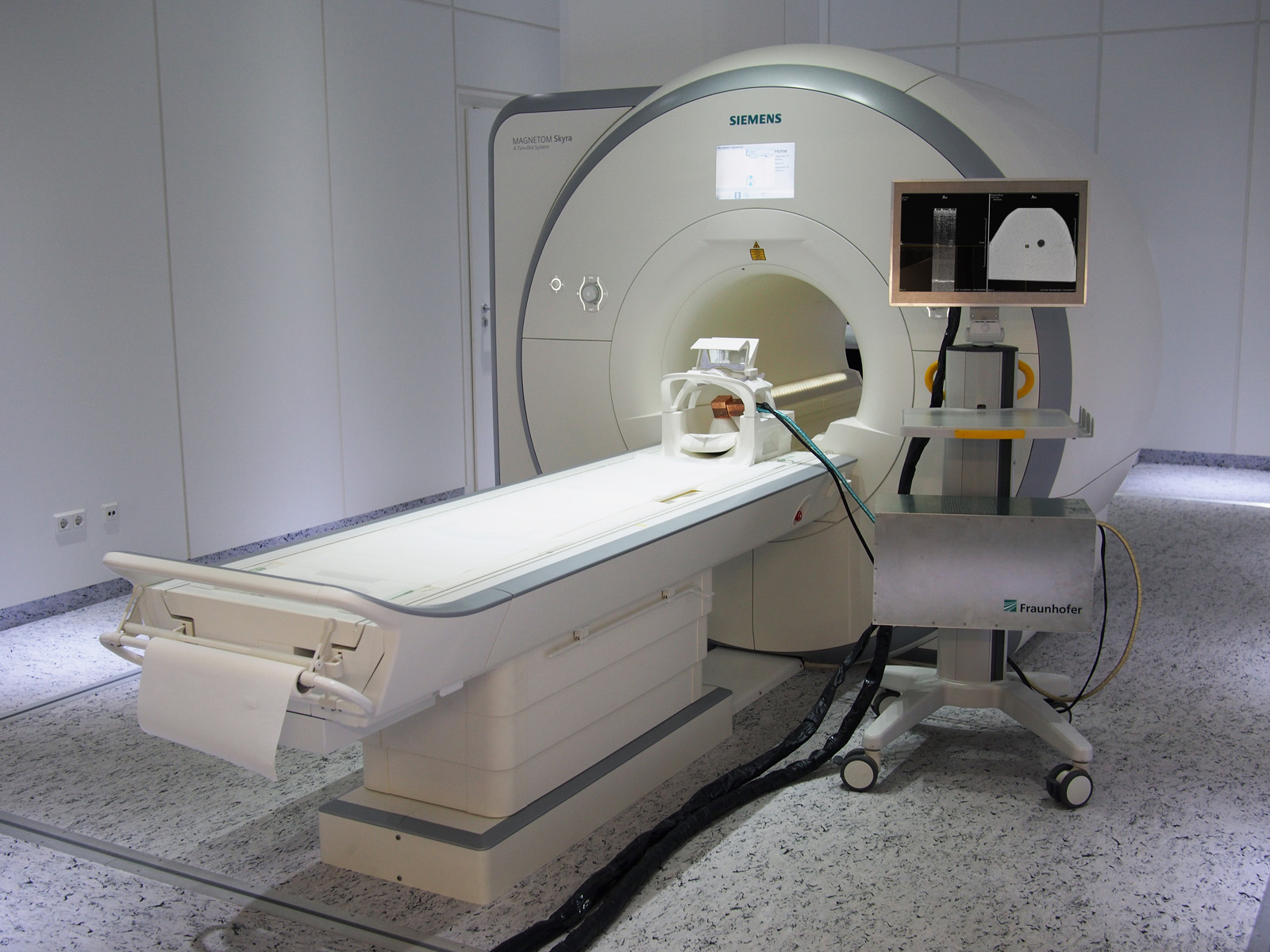
A new technology is in development at Fraunhofer IBMT and Fraunhofer MEVIS, funded by internal and external programs.
It requires just one scan of the patient’s entire chest once at the beginning of the procedure. The subsequent biopsy is guided by ultrasound, while the initial MRI scan is transformed and accurately rendered on the screen. Doctors would have both the live ultrasound scan and a corresponding MR image available to guide the biopsy needle and display exactly where the tumor is located.
The MRI is performed with the patient lying prone and the biopsy while lying on her back. This change of position alters the shape of the patient’s breast and shifts the tumor’s position significantly. To track these changes accurately, ultrasound probes are attached to the patient’s skin to produce two comparable sets of data from ultrasound as well as MR imaging while the patient is in the MRI chamber. While the biopsy is taken in another examination room, the ultrasound probes remain attached, continually recording volume data and tracking the changes of the breast’s shape. Special algorithms analyze these changes and update the MR scan accordingly. The MR image changes analogously to the ultrasound scan. When the biopsy needle is inserted into the breast tissue, a reconciled MRI scan is available along with the ultrasound image on the screen. This greatly improves the accuracy of needle guidance towards the tumor.
To realize this vision, a range of new components were developed. The team of Fraunhofer IBMT and Fraunhofer MEVIS is currently working on putting together a clinical prototype based on these basic technology components.
 Fraunhofer Institute for Digital Medicine MEVIS