Press Releases

Cancel
  • In the 2025/26 school year, the new upper secondary specialization called “Digital Medicine” will launch in Bremen, developed by Fraunhofer MEVIS and the Oberschule am Waller Ring. On January 23 and 24, interested students can learn more about the new offering in the school auditorium at Lange Reihe 81 in Bremen.

    more info
  • Researchers from the Fraunhofer Institute for Digital Medicine MEVIS, the University Medical Center Freiburg, and Stryker Leibinger GmbH & Co. KG have been awarded the Research Prize for Digitalization in Orthopedics and Trauma Surgery 2024 for their publication entitled “Validation of a Finite Element Simulation for Predicting Individual Knee Joint Kinematics.” The award ceremony took place on October 25, 2024, during the German Congress for Orthopedics and Trauma Surgery (DKOU 2024) in Berlin.

    more info
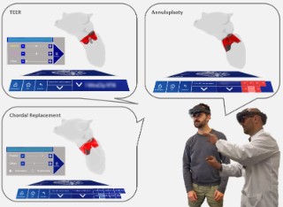
  • Heart valve reconstruction is a highly complex operation that requires a great deal of expertise from surgeons. On the occasion of World Heart Day on September 29, 2024, the Fraunhofer Institute for Digital Medicine MEVIS will present the MINIMAKI assistance system. Based on artificial intelligence methods, it enables interdisciplinary cardiac teams from surgery, cardiology and anesthesia to plan heart valve procedures quickly and optimally using mixed reality.

    more info
  • Data-Efficient Foundation Model for Biomarker Detection

    Artificial Intelligence for Clinical Pathology / September 02, 2024

    The use of Artificial Intelligence (AI) systems shows promise in medicine, where they can be used to detect diseases earlier, improve treatments, and ease staff workloads. But their performance depends on how well the AI is trained. A new multi-task approach to training AI makes it possible to train foundation models quicker and more cost-effectively, with less data. Researchers are turning to this approach to compensate for the shortage of data in medical imaging — and ultimately save lives.

    more info
  • A complete package for minimally invasive tumor therapy

    License agreement and joint development: Fraunhofer MEVIS commences cooperation with Israeli partner for worldwide use of innovative software technology in ultrasound-aided tumor ablation / December 19, 2022

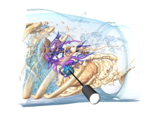
    Image-guided thermal ablation therapy is a patient-friendly, minimally invasive, and cost-effective tumor treatment method. This involves advancing a special needle directly into a tumor using CT and ultrasound to apply heat and destroy it. Until now, interventional radiologists could only estimate the extent to which the applied procedure destroys tumor cells. Hence, thermal ablation is associated with higher local tumor recurrence rates compared with treatment alternatives, causing a real barrier to wide adoption.

    more info
  • MRI scans for early detection of Alzheimer’s disease

    At Fraunhofer MEVIS, a new device update accelerates development of control software / December 20, 2021

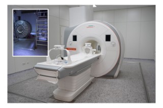
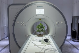
    The pictures show the MR tomograph during and after the upgrade. Most of the device components were replaced except for the superconducting electromagnet.
    © Fraunhofer MEVIS

    Reliable and feasible early detection of Alzheimer’s disease is the goal of DEBBIE, an international joint project under the EU Joint Programme – Neurodegenerative Disease Research (JPND). It is coordinated by the Fraunhofer Institute for Digital Medicine MEVIS in Bremen, which has received national funding from the Federal Ministry of Education and Research (BMBF) for its work. In this project, MRI images shall uncover the extent to which the blood-brain barrier loses function before the first symptoms of Alzheimer’s disease appear. To speed up development, Fraunhofer MEVIS has now enhanced their own MRI scanner to allow much more effective cooperation with clinical partners.

    more info
  • Complications can always occur during operations, sometimes resulting in death. A new project called KIPeriOP aims to minimize the risk of such complications. The project is based on digitized decision guidelines and self-learning algorithms intended to provide reliable risk assessment based on individual patient data. What is the probability that certain complications will occur, and how might they be avoided? The project is coordinated by Prof. Dr. Anja Hennemuth from the Fraunhofer Institute for Digital Medicine MEVIS and Prof. Dr. Patrick Meybohm from the University Hospital of Würzburg. Clinicians from the Asklepios Medical School GmbH, the University Hospital Frankfurt and the Charité – Universitätsmedizin Berlin are involved. They are working together with experts from the fields of AI, user guidance, ethics, and health economics.

    more info
  • novel computer methods, such as those using artificial intelligence and decision support are designed to combine all relevant information, make diagnosis and therapy more efficient, and decrease side effects.
    © Fraunhofer MEVIS

    It took more than six years to plan and build, but now the new building of the Fraunhofer Institute for Digital Medicine MEVIS on the campus of the University of Bremen is completed. To celebrate the inauguration, the institute opens its doors, albeit only virtually due to the current pandemic. After the welcome addresses by Dr. Claudia Schilling, Bremen Senator for Science and Ports, by Alexander Kurz, Executive Vice President Human Resources, Legal Affairs, and IP Management of the Fraunhofer-Gesellschaft, as well as by Prof. Dr. Bernd Scholz-Reiter, President of the University of Bremen, Fraunhofer MEVIS will present a sample of its current research activities. The online event will take place on Friday, June 18, from 2 to 5 pm. The event will be held in German, although the plenary session will be simultaneously interpreted into English, and some presentations will be given in English. Anna Stankiewicz (violin) and Elena Tomarchio (violoncello) from the Konsonanz chamber ensemble provide musical contributions.

    more info
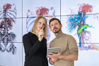
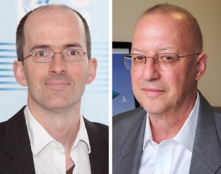
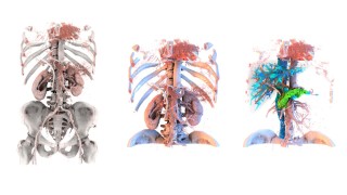
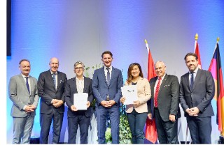
  • Prof. Ron Kikinis, the former director of the Fraunhofer Institute for Digital Medicine MEVIS in Bremen, has accepted a renowned appointment at Harvard Medical School in the United States. Since March 1, 2020, Prof. Horst Hahn has been the sole director of the Institute – for the prior six years, both had acted in dual leadership roles. Kikinis has assumed the B. Leonard Holman Endowed Professor of Radiology at Harvard Medical School. This endowed chair is one of the highest academic distinctions at the prestigious Medical School and is only awarded to researchers who are worldwide leaders in their field.
    more info
  • Patient Bonsai

    November 04, 2019

    During the “STEAM Imaging II” artist residency at Fraunhofer MEVIS, two artists from Singapore develop an interactive installation and participate in a youth workshop.

    more info
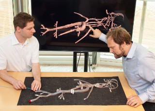
  • Endovascular interventions are an integral part of the medical routine with 6 million procedures done worldwide annually. During the procedure, doctors insert a thin, flexible wire to navigate the catheter into the blood vessels to apply stents or remove blood clots. In order to navigate the catheter precisely through the vessels, patients undergo X-rays during the procedure. One downside is that “patients and doctors are exposed to a considerable amount of radiation,” says Dr. Torben Pätz, mathematician at the Fraunhofer Institute for Digital Medicine MEVIS in Bremen. “In addition, the Xrays merely show a 2D projection instead of a 3D image, which can sometimes impede precise localization of the catheter.” Fraunhofer MEVIS is developing a system called IntelliCath (Intelligent Catheter Navigation) to remedy these problems.
    more info
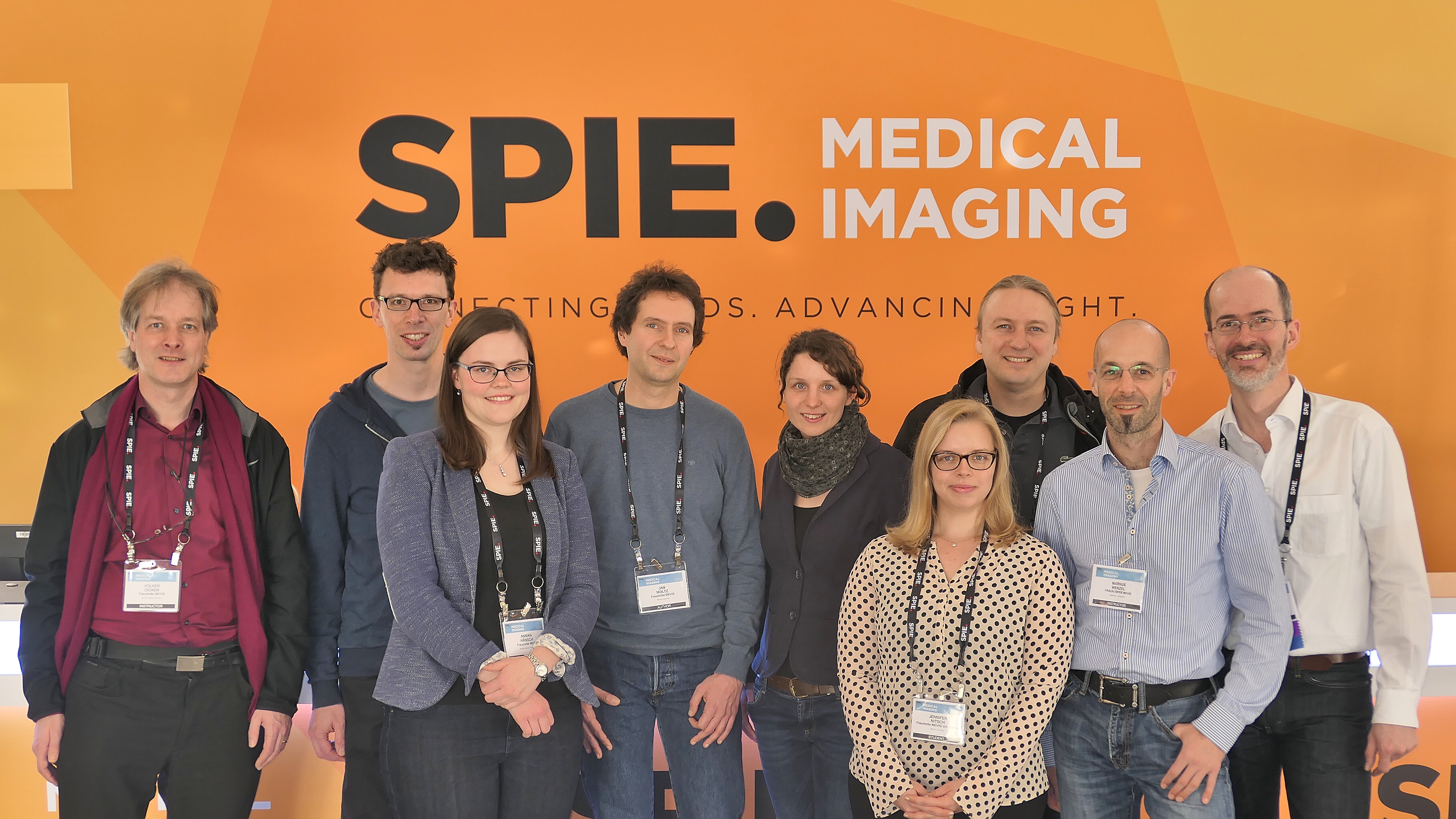
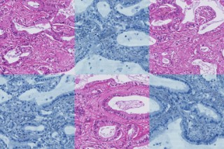
  • Artificial intelligence and adaptive algorithms – in medicine, these are gaining increasing importance. Speaker programs at international conventions also reflect this trend by focusing more and more on possible applications of this new technology. One example is the Medical Imaging conference hosted by the International Society for Optics and Photonics (SPIE), which will take place in San Diego, California, from February 16 to 21. At this renowned convention, the Fraunhofer Institute for Digital Medicine MEVIS will be represented by a number of experts who will provide insight into deep learning.
    more info
  • Das neue Institutsgebäude von Fraunhofer MEVIS
    © Haslob Kruse + Partner

    The Fraunhofer Institute for Medical Image Computing MEVIS is one of the most prestigious research facilities of its kind. At four locations in Bremen, Lübeck, Berlin, and Aachen, the institute develops innovative software systems that support doctors in their daily routine, from heart diagnostics and tumor therapy to big data analysis for large-scale studies. Now, the institute’s main facility in Bremen will move into a new home. The cornerstone for the building will be laid at 11 o’clock on December 5, 2018. Starting in autumn 2020, it will provide a communicative and creative working environment for the MEVIS team.

    more info
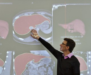
  • Dr. Hans Meine uses computer tomographic images of a liver to describe the contributions that medical computing makes to the analysis of medical images.
    © Fraunhofer MEVIS

    The Fraunhofer MEVIS Institute for Medical Image Computing – in short: Fraunhofer MEVIS – is one of the world’s leading research centers in digital medicine. In order to prepare computer science students for challenges in this area, the institute and the University of Bremen are now cooperating even more closely in teaching. This winter semester saw the introduction of a new study area called Medical Computing in the Faculty of Mathematics / Computer Science.

    more info
  • Inspection of the image-based geometric parameters describing the heart and its function. In patients with mitral valve insufficiency and altered pressure conditions the interplay of these parameters changes.
    © Fraunhofer MEVIS

    One of the world’s most important conferences in the field of computer assisted radiology and surgery, CARS 2018, will take place from June 20 to 23 in Berlin. The Fraunhofer Institute for Medical Image Computing MEVIS in Bremen will make an essential contribution to one of the conference’s key topics: How can minimally invasive heart procedures be performed more effectively and patient-friendly with the help of modern data and image processing algorithms?

    more info
  • A fruitful exchange between art and science
    © Yen Tzu Chang

    On June 5, the Fraunhofer-Gesellschaft will present Taiwanese artist Yen Tzu Chang in the scope of the “Science and Art in Dialog” event series in Berlin. She created a performance entitled “Whose Scalpel” during her artist residency at the Fraunhofer Institute for Medical Image Computing MEVIS. In a subsequent open discussion, Institute Director Horst K. Hahn and Ars Electronica Artistic Director Gerfried Stocker will explore complexity as a term and its possible ramifications for our society.

    more info
  • Four complex, interwoven vascular systems pass through the liver. Surgically removing tumors is therefore often a great challenge. The Fraunhofer Institute for Medical Image Computing MEVIS has developed algorithms that analyze patients‘ imaging data and calculate surgical risks. This makes liver cancer surgery safer and easier to plan.

    more info
  • What effect does a particular cancer medicine or radiation therapy have on patients? To find out, physicians use CT images to determine whether a tumor’s size changes during the course of treatment. In the PANTHER project, a joint team of experts aims at gaining further valuable information from these images. In the future, doctors will be able to find out at an early stage whether a cancer treatment is effective or should be changed. The Fraunhofer Institute for Medical Image Computing MEVIS, with its branches in Bremen and Lübeck, is an essential project partner. This spring, the project team will present the interim results from the first project period.

    more info
  • The AI platform integrates rapid prototyping of clinical applications and immediate inspection of the AI's capabilities on clinical data — while the learning progresses.
    © Fraunhofer MEVIS

    Bit by bit, computer assistants with the ability to learn are finding their way into hospitals and doctors’ practices. They support doctors during diagnosis and therapy planning. Until now, computer scientists played the primary role in designing and developing such ‘deep-learning’ algorithms. The Fraunhofer Institute for Medical Image Computing MEVIS in Bremen, however, is now developing AI systems in close cooperation with physicians. In Chicago on November 26, Fraunhofer MEVIS researchers will present their first promising results at RSNA 2017, the most important radiology congress in the world.

    more info
  • Open-heart art surgery

    August 29, 2017

    Artist Yen Tzu Chang at the MRI labor.
    © Fraunhofer MEVIS

    Yen Tzu Chang is the recipient of the artist residency jointly hosted by Fraunhofer MEVIS and Ars Electronica in the framework of the “European Digital Art and Science Network.” Ars Electronica created the international initiative to enable artists to participate in residencies at prestigious research institutes to connect science with digital art approaches. Chang will present the results of her work during the performance of “Whose Scalpel” at the Ars Electronica Festival in Linz from September 7 to 11.

    more info
  • Dosimetry in the liver
    © Fraunhofer MEVIS

    Radioembolization is a therapy method used for liver tumors that are uncurable with surgery or chemotherapy. Thorough development of an individual planning is important for successful treatment. Within the SIRTOP project, the Fraunhofer Institute for Medical Image Computing MEVIS in Bremen is developing sophisticated computer algorithms to make therapy planning faster and more precise. The research team will present their initial results at several upcoming conferences.

    more info
  • MRI supply regions of specific blood vessels
    © Fraunhofer MEVIS

    When diagnosing strokes and heart diseases or looking at tumors, perfusion magnetic resonance imaging offers a gentler way to capture the blood flow circulation in the organs. However, the method is far from being implemented to its full potential at many clinics. The Fraunhofer Institute for Medical Image Computing MEVIS in Bremen, Germany is organizing a workshop entitled “Measurement of Perfusion and Capillary Exchange” from June 21 to 23 to promote adoption of the method. The event will provide information about its applications and the current state of research. With their joint research alliance, Siemens Healthineers and the Fraunhofer Institute for Medical Image Computing MEVIS will support physicians in finding the right course of therapy for their patients.

    more info
  • Information integration and artificial intelligence for better diagnosis and therapy decisions

    Joint press release of Fraunhofer MEVIS and Siemens Healthineers, Bremen / Erlangen / May 24, 2017

    Volume rendering of vascular tree and metastases in the lung
    © Fraunhofer MEVIS

    Research alliance between Fraunhofer MEVIS and Siemens Healthineers develops decision support systems for physicians based on deep machine learning. With their joint research alliance, Siemens Healthineers and the Fraunhofer Institute for Medical Image Computing MEVIS will support physicians in finding the right course of therapy for their patients.

    more info
  • © Fraunhofer MEVIS

    Focused ultrasound can effectively destroy tumor cells. Until now, this method has only been used for organs such as the prostate and uterus. At the European Congress of Radiology, Fraunhofer researchers will present a method, developed as part of the TRANS-FUSIMO EU project, that enables focused ultrasound treatment of the liver, an organ that moves while breathing. In the future, this could enable treatment of certain liver tumors in a more gentle way.

    more info
  • Bone Metastases
    © Fraunhofer MEVIS

    Physicians have long used visual judgment of medical images to determine the course of cancer treatment. A new program package from Fraunhofer researchers reveals changes in images and facilitates this task using deep learning. The experts will demonstrate this software in Chicago from November 27 to December 2 at RSNA, the world’s largest radiology meeting.

    more info
  • The new robotic assistant requires just five minutes to position the needle.
    © Fraunhofer IPA

    Finding the ideal position for interventional needles – as used in biopsies, for instance – is a difficult and time-consuming process. This can now be performed automatically, using a robotic arm to place a needle guide for the doctor at the optimal insertion point. With robotic assistance, doctors need five minutes to position the needle, as opposed to 30 minutes with conventional techniques. The solution will be shown at the MEDICA trade fair in Düsseldorf from November 14 to 17, 2016 (Hall 10, Booth G05).

    more info
  • Decision Support for Clinicians / 2016

    A Computer Simulation to Spare Children from Heart Surgery

    September 01, 2016

    Simulation Blood Flow Aorta
    © Fraunhofer MEVIS

    Children with congenital heart defects often undergo a battery of strenuous examinations and interventions. In the EU CARDIOPROOF project, Fraunhofer researchers have developed software to simulate certain interventions in advance. Preliminary results point to a reduced need to perform numerous interventions.

    more info
  • meta-realistic medical moving imaging physically based rendering
    © Fraunhofer MEVIS

    On the monitor, a brain spins slowly and can be examined from every angle. Suddenly, some sections start glowing, first on the side and then the entire back of the head. These spectacular images show which brain areas activate when we speak, see, hear, or touch. The method originated at the Fraunhofer Institute for Medical Image Computing MEVIS in Bremen. It uses modern visualization technology called ‘physically based rendering’ in combination with medical image data and enriched with clinically relevant supplementary information. On October 1, the method will premiere in an exhibit at the AUDIOVERSUM Science Center in Innsbruck, Austria.

    more info